 by
by What is an Electroretinogram?
An electroretinogram (ERG) is a diagnostic test that measures the electrical activity generated by cells in the retina of the eye in response to a light stimulus. The ERG reflects the mass response of the retinal cells and provides an objective measure of retinal function.
Recording the ERG
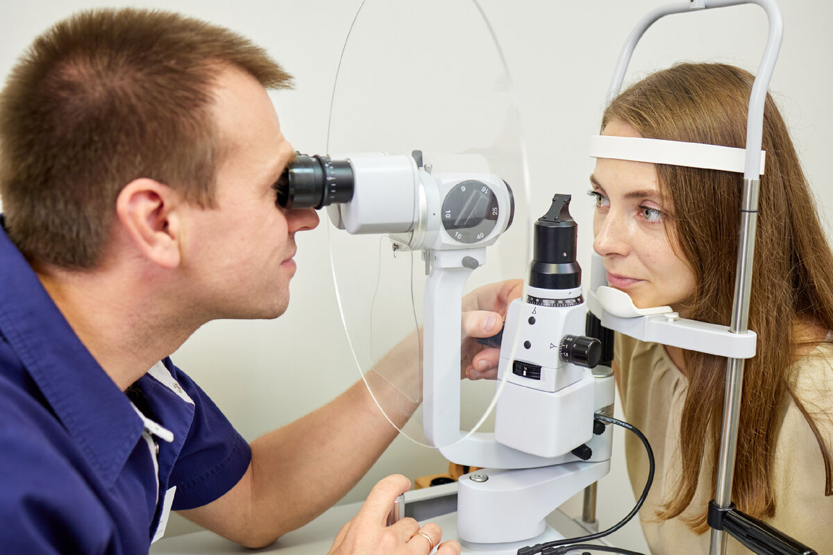
To perform an Global Electroretinogram, special contact lens electrodes are placed on the surface of the eye after application of numbing eye drops. A reference electrode is placed on the skin of the forehead or temple and a ground electrode is attached somewhere else on the body, such as the arm. The electrodes detect the difference in electrical potentials between the retina and other locations on the body.
A light source, typically a photo-stimulator, is then used to flash light into the eye while the electrodes record the retinal response. The signals detected by the electrodes are amplified and displayed on a computer monitor or printed on paper. Different intensities and durations of light flashes are used to assess both the cone and rod systems in the retina. The entire test usually takes 30-60 minutes to complete for each eye.
Types of ERGs
There are different types of ERGs that can be performed to evaluate specific aspects of retinal function:
– Flash ERG: A brief, intense flash of light that monitors overall retinal health. It measures activity from both rods and cones.
– Multifocal ERG: Maps local retinal responses across the visual field by presenting a pattern of hexagons that flash randomly. Useful for detecting local retinal abnormalities.
– Oscillatory potentials: Measures high-frequency wavelets in the b-wave of the flash ERG. Sensitive for detecting inner retinal dysfunction.
– Implicit time: Analyzes the latency of Global Electroretinogram wave components to identify delays that may indicate retinal disease.
– Dark-adapted (rod) ERG: Assesses rod system function by testing in dim red light after dark adaptation.
– Light-adapted (cone) ERG: Evaluates cone system activity after exposing the eye to steady background light to suppress rod function.
Clinical Applications of ERG
The ERG can aid in diagnosing and monitoring many retinal diseases and conditions such as:
– Retinitis pigmentosa – A group of inherited retinal disorders causing progressive vision loss due to photoreceptor cell death. Early ERG changes often precede visual symptoms.
– Diabetic retinopathy – Abnormalities on ERG may predict disease progression in diabetes. Useful for monitoring effectiveness of treatments.
– Macular degeneration – Can detect early involvement of central retina in age-related macular degeneration before it becomes clinically apparent.
– Inherited retinal dystrophies – Different patterns of ERG abnormalities help classify mutations in retinal gene defects.
– Vitamin A deficiency – Low rod and cone functions on ERG indicate nyctalopia and impairments of dark adaptation associated with Avitaminosis A.
– Retinal detachments – An ERG may confirm or rule out a retinal detachment if examination findings are unclear.
– Drug and toxin effects – Useful for evaluating retinal toxicity of pharmaceuticals, recreational drugs, industrial exposures etc. before visual deficits emerge.
Interpreting ERG Results
An ERG performed by an experienced technician and interpreted by a retinal specialist can provide objective data about the health and function of the retina. Abnormal results typically show decreased signal amplitudes or prolonged implicit times compared to age-matched healthy norms. The patterns of abnormalities seen on different ERG tests help localize retinal dysfunction and differentiate disease mechanisms. A normal ERG does not completely rule out early retinal disease but implies overall retinal health at that time. Repeat testing can track changes over time or gauge treatment responses in progressive retinal conditions. The ERG remains a valuable diagnostic and monitoring tool for comprehensive evaluation of retinal function.
*Note:
1. Source: Coherent Market Insights, Public sources, Desk research
2. We have leveraged AI tools to mine information and compile it.