
 by
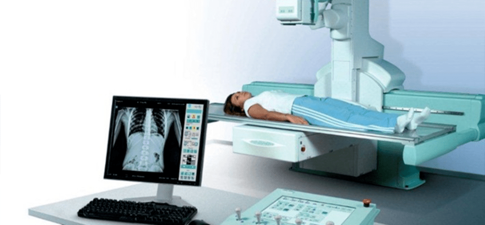
by Fluoroscopy refers to a type of medical imaging that shows a continuous X-ray image on a monitor, much like an analog X-ray machine. Traditional fluoroscopy uses a image intensifier and TV camera to convert the X-rays into a video image in real-time. However, with the rise of digital technology, newer digital fluoroscopy systems are replacing the older analog versions.
Advantages of Digital Fluoroscopy
Digital Fluoroscopy System offer several key advantages over traditional analog fluoroscopy:
Higher Image Quality: Digital systems produce crystal clear images with high resolution and contrast compared to the grainy images from analog systems. Doctors can see more detail which helps improve diagnostic accuracy.
Low Radiation Exposure: By controlling technoligal parameters like pulse rate, current and tomography, digital systems minimize unnecessary radiation exposure to both patient and medical staff. Advanced processing algorithms help enhance image quality while using up to 70% less radiation.
Image Storage and Retrieval: Digital images can be stored indefinitely on PACS (picture archiving and communication systems) for future reference and comparison. Doctors can easily retrieve prior scans and studies for patients. Traditional analog images had to be developed from film and stored in physical archives.
Telemedicine Capabilities: Digital images allow remote consultation and expertise to be utilized via secure data streaming. Radiologists and surgeons can discuss cases over internet with peers in other locations and guide procedures remotely. This enhances treatment quality in rural areas.
Multi-Modality Integration: Images from digital fluoroscopy, CT, MRI, ultrasound etc can all be reviewed and analyzed together seamlessly on electronic monitors. This aids comprehensive diagnosis by evaluating multiple tests at one place.
Cost Savings: Digital Fluoroscopy System are now comparable in price to older analog ones and have minimal maintenance costs. Film and chemicals are not needed which reduces running expenses over the long term. Overall total cost of ownership is lower for digital options currently.
Components of a Digital Fluoroscopy System
A digital fluoroscopy system typically consists of the following key technological components:
Fluoroscopy X-Ray Tube: Produces pulsed beams of X-rays which pass through the patient’s body and strike the detector. Tube positioning and angles can be adjusted and controlled remotely.
Digital X-Ray Detector: Instead of an image intensifier, it uses a flat panel digital detector like CCD (charge-coupled device) or CMOS (complementary metal–oxide–semiconductor) sensor. These convert x-ray photons into electric signals representing the radiographic image.
Image Processing Computer: Performs tasks like image enhancement, digital subtraction of before and after images, video recording/storage, application of controls like magnification etc. Advanced computers provide high-speed real-time image processing.
Medical Grade Monitors: Multiple high-resolution LCD or LED displays show live fluoroscopic video and stored images/scans to medical staff for guidance during procedures and evaluations.
Control Room: This encloses all equipment and electronic components away from radiation in a shielded space. Technologists operate the system via network cables and wireless remotes outside patient rooms ensuring safety.
Applications of Digital Fluoroscopy
Some common medical procedures where digital fluoroscopy plays a vital role include:
Cardiology: Visualization of heart and blood vessels during angiograms, stent placements, valvuloplasties, ablations etc. Movement is clearly tracked.
Gastroenterology: Examinations of esophagus, stomach and intestines like upper GI studies, balloon dilatations, biopsies, endovascular treatments etc.
Urology: Procedures involving urinary tract like pyelograms to study kidneys/ureters and urethrograms to examine urethra are easily performed.
Orthopedics: Real-time imaging of joints, muscles and bones aids minimally invasive surgeries of spine, hips, knees etc. Percutaneous injection therapies too.
Interventional Radiology: Image-guided placement of tubes, drains, vascular access devices and other interventional tools inside the body precisely.
The Future of Digital Fluoroscopy
As digital fluoroscopy becomes more sophisticated, new ways of enhancing its capabilities are being researched. Some likely future developments include:
Higher Frame Rates: Real-time imaging of fast moving anatomy like heart will be clearer with faster acquisition frame rates possibly exceeding 60 fps in future systems.
3D/4D Imaging: Using tomosynthesis or other techniques to reconstruct 3D and 4D (real-time 3D) volumetric images from multiple projection angles can offer new clinical benefits over 2D views alone.
Fluoroscopy Fusion: Advanced registration of other scanned images (CT/MRI/PET etc) directly onto live fluoro videos may improve procedural outcomes through augmented reality guidance.
Lower Dose Radiation: New detector designs and improved image processing aim to bring radiation levels down to as low as reasonably achievable (ALARA) principle through dose reduction techniques.
Remote Access: 5G networks might enable high-quality real-time streaming of digital fluoroscopy to any internet-connected device, enabling teleconsultations from home.
AI Integration: Application of machine learning algorithms for automatic image analysis, anatomical structure recognition, procedure quality assessment etc could transform the usage of digital fluoroscopy.
In summary, digital fluoroscopy systems have revitalized this important imaging technique through technological upgrades that are enhancing medical care via improved accuracy, real-time guidance and radiation safety. Further research ensures its continued utility well into the future.
*Note:
1. Source: Coherent Market Insights, Public sources, Desk research
2. We have leveraged AI tools to mine information and compile it

About Author - Vaagisha Singh
Vaagisha brings over three years of expertise as a content editor in the market research domain. Originally a creative writer, she discovered her passion for editing, combining her flair for writing with a meticulous eye for detail. Her ability to craft and refine compelling content makes her an invaluable asset in delivering polished and engaging write-ups. LinkedIn


